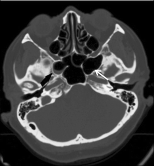
90 Amazing Soft Tissue Sphenoid Sinus
Soft tissue sphenoid sinus As they are not so prominent in this case it is easy to identify and spot them. CT nicely demonstrates the bone destruction and some of the soft tissue involvement.
 Orbital Lamina Of Ethmoid Bone Lamina Papyracea Its Name Lamina Papyracea Is An Appropriate Description Dental Hygiene School Rectus Muscle Sphenoid Bone
This particular Doctor saw a soft tissue mass in the sphenoid sinus area.

Soft tissue sphenoid sinus. Fluid right middle ear cavity and right mastoid air cells suggesting an active inflammatory process. Coronal reformatted image in a soft-tissue window from a noncontrast CT scan shows a soft-tissue mass centered on the sphenoid sinus with dehiscence of the planum sphenoidale arrow. According to his expertise he said the bones that surround the dark area hole of the sphenoid sinus - the one on the left side brokedisinergrated whatever and the soft tissue spilled into that area. Soft tissue sphenoid sinus
It usually lies anteroinferior to the sella. There is opacification of the sphenoid sinus with destruction of and osteopenia of the sphenoid bone. It is difficult to identify diseases in this part because of its location. Soft tissue sphenoid sinus
In front of the sphenoid sinuses are the ethmoid sinuses. 1 doctor answer 2 doctors weighed in My MRI reported. In severe cases in general the sinuses are completely obliterated and remodeling of sinonasal bones is common. Soft tissue sphenoid sinus
Axelsson and Jensen found that when plain radiography was performed on 300 patients specifically referred with a clinical diagnosis of sinusitis 68 had abnormal studies but none showed abnormalities in the sphenoid sinus. Silvers They act as airbags to the brain. Sinonasal polyposis is usually characterized on CT as soft tissue density polypoid masses within the nasal cavity and paranasal sinuses. Soft tissue sphenoid sinus
The image on the right is more cranial. The head would weigh more and be exhausting to carry around Finally says Dr. Sphenoid sinuses are just behind the skull above the nasopharynx and just below the pituitary gland. Soft tissue sphenoid sinus
The rest of the sinuses are. It could indicate chronic infection as well. The sphenoid sinus is situated at the center of the skull. Soft tissue sphenoid sinus
The nasopharyngeal soft tissue and the adenoids are also well visualized. Sphenoid sinus mucosal thickening occurs when the lining of the sinus cavities swell. However the sphenoid sinus is obscured. Soft tissue sphenoid sinus
The CT scan exhibited a non-enhancing 3 5 cm soft-tissue mass centered in the sphenoid sinus and extending into the left ethmoid sinuses and the pterygopalatine fossa as well as the middle cranial fossa Figure 1. MR better images the contents of the cephalocele showing CSF and herniated meninges andor brain tissue. Soft tissue densities in the normally air filled mastoid middle ear and sinus cavities indicates there is some type of chronic long term inflammation in those areas. Soft tissue sphenoid sinus
Isolated sphenoid sinus disease is unusual and one should at least consider the possibility of a cephalocele with a rounded soft tissue density in the sphenoid sinus. Large soft tissue mass in the masticator space asterisk. Here we explore what causes sphenoid sinusitis and how to deal with it. Soft tissue sphenoid sinus
Sphenoid sinusitis is an inflammation-related condition that can create uncomfortable symptoms and headache pain. CT is excellent in demonstrating the bony skull base defect. In the lateral view the sphenoid and frontal sinuses are visualized. Soft tissue sphenoid sinus
Wha does this mean soft tissue densities within mastoid cells middle ear cavity and sphenoid sinus and calcific density in brain paren occipital reg. Surrounding circumferential hypoattenuating mucosal thickening is usually present indicating chronic sinusitis. 13 CT shows fungus ball as a high-density mass with calcification. Soft tissue sphenoid sinus
If an unfortunate trauma occurs to the face the sinuses collapse protecting the posterior vital. The maxillary sinuses are most commonly affected followed by the sphenoid frontal and ethmoid sinuses. A modified basilar view a submental vertex view may be a useful. Soft tissue sphenoid sinus
Cysts filled with mucous can form in the cavities when a gland becomes obstructed and swells. The sphenoid sinuses are paired spaces formed within the body of the sphenoid bone communicating with the roof of the nasal cavity via the sphenoethmoidal recessin its anterior wall. Many vulnerable structures surround this sinus for example Proetz mentioned the dura mater cranial nerves III IV V1 V2 and VI optic nerve and chiasm internal carotid artery cavernous sinus pituitary gland sphenopalatine ganglion sphenopalatine artery and pterygoid canal 1. Soft tissue sphenoid sinus
Hi Soft Tissue Attenuation the sinus is made of bone hard tissue but its linned by a mucus membrane soft tissue and attenuation is a radiological term meaning a change from base line indicate increased water content most likely due to an inflammation. Prior infections can cause swelling and swelling can also lead to an infection. Any bulge of soft tissue that is seen in the sinus is abnormal. Soft tissue sphenoid sinus
The next function is that they aerate the skull imagine if sinuses were solid bone or full of soft tissue. The two sinuses are separated by a septum which may or may not be in the midline. The anterior ethmoid air cells are also seen. Soft tissue sphenoid sinus
 Isolated Sinusitis Of Lateral Recesses Of Sphenoid Sinus Springerlink
 Heterogeneous High Density Polypoidal Mucosal Thickening Involving Right Maxillary Sinus Antrum Extending Thr Diagnostic Imaging Sinus Drainage Maxillary Sinus
 Ct Findings The Left Sphenoid Sinus Shows A Soft Tissue Shadow Without Download Scientific Diagram
 A 55 Year Old Woman With Ngh In The Sphenoid Sinus Axial And Sagittal Download Scientific Diagram
 Acute And Chronic Sinusitis And Allergies
 Functional Endoscopic Sinus Surgery Overview Preparation Technique Sinus Surgery Sinusitis Paranasal Sinuses
 Anatomy Physiology Of Nose Nasal And Paranasal Sinus Paranasal Sinuses Sinus Cavities Sinusitis
 Maxillary Ethmoid Spenoid Frontal Sinuses Sinusitis Respiratory System Anatomy Body Systems
 Axial Ct Showing Soft Tissue Masses In The Right Ethmoid Sinus With Download Scientific Diagram
 Juvenile Nasopharyngeal Angiofibroma Typically A Lobulated Non Encapsulated Soft Tissue Mass Is Demonstrated Centred On Th Radiology Pet Ct Nasal Obstruction
Sphenoidal Sinus Osteomyelitis A Fat Suppressed T2 Image In The Axial Download Scientific Diagram
 Image Result For Lateral Wall Of Nose Nose Diagram Nasal Cavity Nose
 Pin By Dorlecia Blanks On Back To School Nasal Cavity Sinus Cavities Sinusitis
 Sphenoid Sinus Neuroendocrine Carcinoma A Axial Ct Bone Windows Download Scientific Diagram
 This Image Shows The Sagittal Section Of The Bones That Comprise The Nasal Cavity Nasal Septum Nasal Cavity Anatomy And Physiology


